Good long term results for tissue grafts?

Methods:

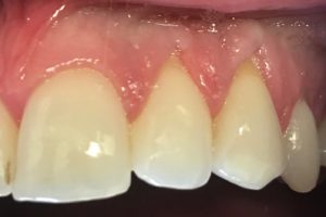
Forty-seven patients with 64 sites with lack of attached gingiva were treated with free gingival grafts and 64 control sites were left untreated.  Patients were recalled every 4 to 6 months.  Gingival recession depth, keratinized tissue width, and probing depth were measured at baseline and extending to the follow up period (18 to 35 years)

Results:

At the end of the follow up period, 83% of the 64 treated sites showed recession reduction whereas 48% of the 64 untreated sites experienced increase in recession. Treated sites ended with gingival margin 1.7 mm more coronal and keratinized tissue 3.3 mm wider than untreated sites.

Conclusions:

Sites treated with gingival augmentation procedures resulted in coronal displacement of the gingival margin and reduced recession, whereas the contralateral untreated sites showed a tendency for increased recession and a tendency to develop new recession during the 18 to 3 year follow up.

For more information please refer to:

Journal of Periodontology 2009 Sep;80(9):1399-405

Periodontal conditions of sites treated with gingival augmentation surgery compared to untreated contralateral homologous sites: a 10- to 27-year long-term study.

Agudio G1, Nieri M, Rotundo R, Franceschi D, Cortellini P, Pini Prato GP.


Platelet-rich fibrin (PRF) to treat periodontal intrabony defects?

Methods:

36 intrabony defects in 36 individual patients were treated with either demineralized freeze-dried bone allograft (DFDBA) or platelet-rich fibrin (PRF).  Data was collected at baseline and at 6 month follow up and included radiographic bone fill and changes in clinical attachment levels (CAL).

Results:

Both treatment groups had significant gain in clinical attachment levels and bone fill, with no significant difference between groups.  Mean CAL gain for DFDBA was 1.16 ± 1.33 mm and mean radiographic bone fill was 1.14 ± 0.88 mm.  PRF had a mean CAL gain of 1.03 ± 0.86 and mean radiographic bone fill of 1.10 ± 1.01 mm.

Conclusions:

Intrabony defects treated with either DFDBA or PRF had a significant gain in CAL as well as bone fill after 6 months of healing and there was no significant difference between the two materials.

For more information please refer to:

Journal of Periodontology, November 2016, Vol. 87, No. 11

Clinical and Radiographic Evaluation of Demineralized Freeze-Dried Bone Allograft Versus Platelet-Rich Fibrin for the Treatment of Periodontal Intrabony Defects in Humans

Jane K. Chadwick, Michael P. Mills,  and Brian L. Mealey

“Growth and Profit for Your Practice”- a Full Day CE Seminar on Sunday, November 20th, 2016 at the Trump International Hotel, 325 Bay Street, Toronto.

This course will help you transform your practice, attract more patients, and increase profitability.

Course Objectives:

How to attract new patients and master maintenance retention strategies.

How to communicate treatment plans for greater case acceptance.

Learn to do online keyword research for your dental practice and the most effective keywords for practice growth.

How to check your keyword ranking and how to gather information on other practices in your area.

Learn how emotion and psychology influence practice growth decisions and how to manage downside risk.

Learn tax planning to keep more in pocket and how to credit proof and litigation proof your assets.

How to create a raving fan base by giving patients a VIP experience.

How to engage your staff and get them to take ownership of their part in your success.

Learn how not to overspend on dental equipment by comparing actual product specifications.

Full Day Seminar Eligible for 6 RCDSO Credits (Category 3)

COURSE FEE: $595 + 13% HST. Breakfast and lunch included. Early Registration/ AGD Discount: $525 + 13% HST if you register by October 21, 2016

Download the registration form at SCIENCEOFIMPLANTS.COM

For more course information or to register by phone, please contact Sarah Ellery at (519) 278-6553
growthandprofit

Are Long Terms Results for Collagen Matrix Comparable to Autogenous Tissue for Root Coverage?

IMG_0950(05-20-19-15-01)_20160520

Methods:

Collagen matrix used with a coronally advanced flap was compared to a connective tissue graft used with a coronally advanced flap at 6 months and 5 years for root coverage achieved.

Results:

Seventeen patients were available for the 5-year recall.  Mean root coverage between 6 months and 5 years changed from 89.5% to 77.6% for collagen matrix and 97.5% to 95.5% for connective tissue.  There was no significant difference between the therapies for changes in root coverage, keratinized tissue width, and probing depth.  Patient satisfaction was similar for both therapies.

Conclusions:

Collagen matrix is a viable long term alternative to traditional autogenous connective tissue when used in conjunction with a coronally advanced flap for root coverage procedures.

Dr. G’s comments:

There is a trend for slightly better root coverage with autogenous tissue, but patients seem to prefer the lower morbidity of an allograft.

For more information please refer to:

J Periodontol. 2016 Mar;87(3):221-7

Long-Term Results Comparing Xenogeneic Collagen Matrix and Autogenous Connective Tissue Grafts With Coronally Advanced Flaps for Treatment of Dehiscence-Type Recession Defects.

McGuire MK, Scheyer ET

Does Suture Removal Time Affect Root Coverage Outcome?

SCIENCE OF IMPLANTS May 8 2016
Methods:
A meta-analysis of randomized clinical trials that assessed single tooth class I or II recession defects treated surgically with a coronally advanced flap. Early (less than 10 days post-op) and late (more than 10 days post-op) suture removal were compared in terms of differences in complete root coverage outcome.
Results:
Data from seventeen studies comprising of 325 single recession defects revealed a superior proportion of sites exhibiting complete root coverage when sutures where removed more than 10 days post-operatively.  No significant difference in outcomes between absorbable and non-absorbable sutures when they were removed more than 10 days after surgery.
Conclusions:
Early suture removal (less than 10 days post-op) can hinder complete root coverage outcomes in singe tooth recession defects treated with a coronally advanced flap.
Dr. Gebrael’s comments: 
Modern 5-0 polypropylene sutures have excellent tensile characteristics and are comfortable enough for us to delay suture removal until at least one month post-op.  This allows a mature attachment to form and increases the odds for complete root coverage.
For more information please refer to:    J Periodontol. 2016 Feb;87(2):148-55
The Effect of Suturing Protocols on Coronally Advanced Flap Root-Coverage Outcomes: A Meta-Analysis
Tatakis DN, Chambrone L

Is Root Coverage Effective for Dentin Sensitivity?

Methods

A systematic review of the literature regarding the efficacy of surgical root coverage at reducing cervical dentin hypersensitivity in cases of gingival recession yielded nine randomized clinical trials suitable for analysis. The primary outcome included changes in pain/hypersensitivity symptoms before and after surgical treatment.

Results:

A reduction in cervical dentin hypersensitivity was reported in all studies. The mean percentage of decreased hypersensitivity was 77.83% with follow-up ranging from 3 to 30 months. The size of the effect was small in some studies and the risk of bias was considered high. None of the studies performed a correlation between hypersensitivity and percentage of root coverage.

Conclusions:

There is not enough evidence to conclude that surgical root coverage procedures can predictably reduce sensitivity. More robust studies are needed to determine the effectiveness of surgical root coverage for the treatment of cervical dentin sensitivity.

Dr. G’s comments:

There are many painful and unpleasant consequences to dentin sensitivity including food and drink restrictions. Root coverage procedure appear to decrease sensitivity but the evidence is weak.

FOR MORE INFORMATION PLEASE REFER TO :

J Periodontol. 2013 Mar;84(3):295-306

Is surgical root coverage effective for the treatment of cervical dentin hypersensitivity? A systematic review.

Douglas de Oliveira DW1, Oliveira-Ferreira F, Flecha OD, Gonçalves PF.

How Successful is Peri-Implantitis Treatment?


Methods:

A retrospective study on 382 implants with peri-implantitis in 150 patients. Peri-implantitis was defined as pockets ≥ 5 mm, bleeding on probing or suppuration, and the presence of radiographic bone loss ≥ 3 mm or at least three threads of the implant.

Results:

The mean time between implant installation and peri-implantitis was 6.4 years. Periodontal flap surgery was the most common treatment and was performed in 47% of cases. Regenerative treatment was the treatment choice in 20% of cases. Successful treatment was defined as absence of bleeding on probing and/or suppuration and a probing depth of < 4 mm and this was achieved in 69% of patients and for 82% of implants with a mean follow-up time of 26 ± 20 months.

Conclusions:

High success rates for peri-implantitis treatment with a mean follow up of ≈ 2 years. Therapy was less successful for patients with severe periodontitis, severe mean marginal bone loss around the implants, poor oral hygiene, and low compliance.

For more information please refer to:

Treatment Outcome in Patients With Peri-Implantitis in a Periodontal Clinic: A Retrospective Study

Maria Lagervall and Leif E. Jansson

Journal of Periodontology

October 2013, Vol. 84, No. 10

Do block graft complications lead to treatment failure?

Methods:

Implant supported restorations were placed in 137 atrophic ridges that were augmented with cancellous block-bone allografts. Recipient site complications associated with block grafting were recorded.

Results:

Total block-graft failure occurred in 7% of cases and partial block graft failure occurred in 8% of cases. The implant failure rate was 4.4%. Soft tissue complications included membrane exposure (42 of 137), incision line opening (41 of 137), and perforation of the mucosa of the grafted bone (19 of 137). A higher rate of complications was noted in the mandible.

Conclusions:

Soft tissue complications did not necessarily result in total loss of cancellous block allograft. Total bone-block failure (8% of cases) and implant failure (4.4% of cases) were uncommon with block grafting although approximately 30% of patients experienced adverse events.

for more information please refer to :

J Periodontol. 2010 Dec;81(12):1759-64

Analysis of complications following augmentation with cancellous block allografts.

Chaushu G1, Mardinger O, Peleg M, Ghelfan O, Nissan J.

Does Timing of Restoration Affect Implant Marginal Bone Loss?


METHODS:


A meta-analysis was performed on eleven studies that compared marginal bone loss between implants restored with the following protocols: Immediate restoration/loading; early loading; conventional loading 


RESULTS:


 After adjusting for implant placement levels, no significant difference was found when comparing marginal bone loss for four scenarios: 

  1. immediate restoration/loading + delayed placement versus conventional loading + delayed placement.
  2. immediate restoration + delayed placement versus early loading + delayed placement.  
  3. early loading + delayed placement versus conventional loading + delayed placement.
  4. immediate loading + immediate placement versus conventional loading + immediate placement.

CONCLUSIONS:

The timing of restoration does not have an effect on implant marginal bone loss according to this meta-analysis when comparing the clinical scenarios outlined above.


FOR MORE INFORMATION PLEASE REFER TO:


Effect of the timing of restoration on implant marginal bone loss: a systematic review.


Suarez F, Chan HL, Monje A, Galindo-Moreno P, Wang HL.


J Periodontol. 2013 Feb;84(2):159-69.


Does Sinus Membrane Perforation Affect Implant Survival?

Methods:

Retrospective data was obtained from 23 patients who had undergone sinus augmentation procedures with a total of 40 treated sinuses.  Sinuses were grafted with mineralized cancellous bone allograft, anorganic bovine bone matrix, or biphasic calcium phosphate.  There were 15 perforated sinuses which were repaired with an absorbable collagen membrane.  Histologic cores were taken from all treated sinuses 26 to 32 weeks after surgery.  

Results: 

Average percentage of vital bone was 26.3% ± 6.3% in the repaired sinuses versus 19.1% ± 6.3% in the non-perforated sinuses.  The implant success rate was 100% in the repaired sinuses versus 95.5% in the non-perforated sinuses.  There was no statistically significant difference in the failure rates.

Conclusions:

There was greater vital bone formation in the repaired sinuses compared to the non-perforated sinuses after augmentation.  When properly repaired, maxillary sinus membrane perforations do not appear to adversely affect vital bone formation or implant survival.

Dr. Gebrael’s comments:

Interesting enough we see more vital bone formation in the repaired sinuses after grafting.  The authors of the study indicate this may be due to the membrane over the perforation acting as a second barrier that prevents soft tissue migration and immobilizes the graft particles.

For more information please refer to:

Effect of maxillary sinus membrane perforation on vital bone formation and implant survival: a retrospective study.

Froum SJ, Khouly I, Favero G, Cho SC.

J Periodontol. 2013 Aug;84(8):1094-9